The Silent and Recurrent Cause of Premature Birth: Cervical Insufficiency
Premature births and pregnancy losses are traumatic and devastating experiences for mothers, fathers, and the entire family. Premature babies have difficulty surviving. For those who survive, there are risks of lifelong disability and need for assistance.
Premature births and pregnancy losses are traumatic and devastating experiences for mothers, fathers, and the entire family. Premature babies have difficulty surviving. For those who survive, there are risks of lifelong disability and need for assistance. For these reasons, the medical world is making intense efforts to investigate the causes of premature births and prevent this condition. Despite all scientific and technological advances today, the risk of premature birth unfortunately cannot be significantly reduced, and approximately 10 out of every 100 pregnancies occur as premature births.
The cause of premature births is not singular. Sometimes early contractions and labor pains begin, sometimes due to congenital anomalies of the uterus and the uterus not reaching the volume to carry the baby, and sometimes doctors are forced to perform premature delivery for the mother and baby.
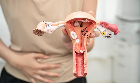
The organ that carries the baby in the mother's womb during pregnancy is called the uterus. The uterus essentially consists of two parts: the uterine body (corpus) and the cervix. During pregnancy, the uterine body expands as the pregnancy progresses and the baby grows, providing space for the growing baby to remain inside. The cervix is at the bottom of the uterus and serves as a barrier during pregnancy so that the mass formed in the uterus due to the growing baby, the baby's waters, and the baby's companion (placenta) can continue to remain inside the uterus. Thanks to this barrier function that the cervix must perform throughout pregnancy, the pregnancy material (baby, placenta, and waters) remains inside the uterus and pregnancy continues. When the cervix shows weakness in performing this barrier function, the cervix shortens, thins, and opens before pregnancy reaches full term. In this case, the pregnancy material inside the uterus is expelled even though there are no labor pains. This condition that occurs due to weakness in the cervix is called cervical insufficiency.
Pregnancy losses due to cervical insufficiency usually occur during the second trimester of pregnancy or at the beginning of the third trimester. Even though there are no uterine contractions (labor pains), the cervix spontaneously shortens, thins, and opens. As a result, the pregnancy material inside the uterus is expelled and pregnancy loss occurs.
Normally, the cervix is strong enough to keep the mass of the baby and its appendages settled inside the uterus for 40 weeks. Why does the cervix remain weak and lead to pregnancy loss?
Main Causes of Cervical Insufficiency
- Surgical procedures performed on the cervix: Conization (removal of precancerous lesions of the cervix)
- LEEP: Similar to conization, removal of tissue from the cervix
- Trauma and tears: Cervical weakness due to causes such as delivery or curettage
- Congenitally weak cervix
How Is Cervical Insufficiency Diagnosed?
Cervical insufficiency is usually understood from the patient's history. Patients report that in their previous pregnancies, the cervix opened painlessly in the middle of the second trimester or at the beginning of the third trimester. This situation has occurred repeatedly in cases where no intervention was made. If the patient does not have such a history, then during pregnancy examinations, it can be detected through transvaginal ultrasonography that the cervix has shortened and even taken on a funnel-like shape. Some cases may come to us with the cervix already opened and the baby's membranes visible in the vagina.
Can Cervical Insufficiency Be Treated?
Cervical insufficiency is a condition that can be treated and has a high treatment success rate.
If cervical insufficiency is known before a planned pregnancy, it is possible to treat it with medications to be used throughout pregnancy and a purse-string suture (cerclage) placed on the cervix at the 12th week of pregnancy, and the treatment success rate is over ninety percent.
If cervical insufficiency is not known beforehand, cervical insufficiency can be diagnosed by evaluating the cervix with transvaginal ultrasonography during pregnancy check-ups. Especially measuring the cervix shorter than 25 mm between the 24th-28th weeks of pregnancy, seeing funneling in the cervix suggests cervical insufficiency, and in this case, medications or placing a purse-string suture (cerclage) on the cervix works in treatment. However, the success rate of cerclage surgery performed in emergency situations is around 75 percent, which is lower compared to planned cerclage performed at the 12th week of pregnancy.
Conclusion
Cervical insufficiency, which can cause premature birth and related death or disability conditions, is an important problem. Careful investigation of the pregnant woman's history and ultrasonographic evaluation of the cervix for cervical insufficiency during pregnancy contributes to the diagnosis of the problem and provides us with an opportunity for intervention.