What is Myoma?
Myomas are the most common tumors of the female genital system and uterus. Myomas are detected in one in every three to two women of childbearing age. While many myomas are diagnosed incidentally, a significant number are diagnosed during gynecological examinations by physicians who are referred due to complaints.
Why Do Myomas Form?
Myomas are caused by excessive proliferation of smooth muscle cells in the uterus or vessels within the uterus. The most critical hormone in the formation of myomas is the female hormone known as estrogen. While myomas are not seen in the pre-puberty period when there is no estrogen hormone, myomas begin to shrink when the estrogen hormone decreases during menopause. Since the estrogen hormone is higher than pre-pregnancy levels during pregnancy, myomas grow rapidly during pregnancy.
What Kind of Complaints Do Myomas Cause?
Myomas seen in the uterine wall, depending on their size and location, sometimes do not cause any complaints, but sometimes they can be the cause of many problems such as increased menstrual bleeding, intermenstrual bleeding or continuous menstrual bleeding, pain in the groin and during sexual activity, frequent urination by pressing on surrounding organs, constipation, inability to conceive (infertility), pregnancy loss, i.e. miscarriage or premature birth. Myomas that grow outside the uterus sometimes grow silently and women do not think of myoma as the cause of abdominal swelling and think that they are gaining weight and they waste time between dietitians and gyms. Women with excessive bleeding due to myoma are often weak and talk about anemia, low ferritin and iron. However, the real reason is anemia caused by bleeding due to myomas and the weakness related to it. In addition, the presence of large and numerous myomas creates an additional load on the heart for feeding the uterus growing with these myomas. All these are the weakness, getting tired easily, decreased exercise capacity, etc. seen in women with myomas. may indicate myomas as the cause of the complaints.
How is Myoma Diagnosed?
In women who have the above complaints or who have no complaints and undergo gynecological examination, the presence of a uterus that is larger than normal and has irregular edges during manual examination raises suspicion of the presence of myoma. When an examination is performed with ultrasonography, which is an indispensable diagnostic tool for gynecologists and obstetricians today, myomas in the uterus (growing inside, on the wall or outside) can be easily recognized. In many cases, myomas can be diagnosed incidentally in lower abdominal ultrasonography, MRI or CT examinations performed on women who apply to the hospital for any reason.
One of the most important points to consider when diagnosing myoma is whether the real diagnosis is myoma. Because in a small number of masses that are evaluated as myomas by ultrasound in the uterus (less than one percent), there is a risk of cancer cells in these masses, even though ultrasound or MRI images suggest myoma. The presence of cancer cells in masses that appear to be myomas often turns out to be sarcoma (leiomyosarcoma: LMS). The only way to definitively understand whether there are cancer cells in masses that appear to be myomas by ultrasound is to have these masses removed and sent for pathological examination. Pathological examination can determine whether the mass is really a myoma or a sarcoma. While some women think that they have myomas with ultrasound imaging and that they are being followed up, it may be too late if cancer cells are found in myomas that are removed with surgery. Therefore, it should always be kept in mind that there may be cancer cells, although rare, in masses that are thought to be myomas by imaging methods.
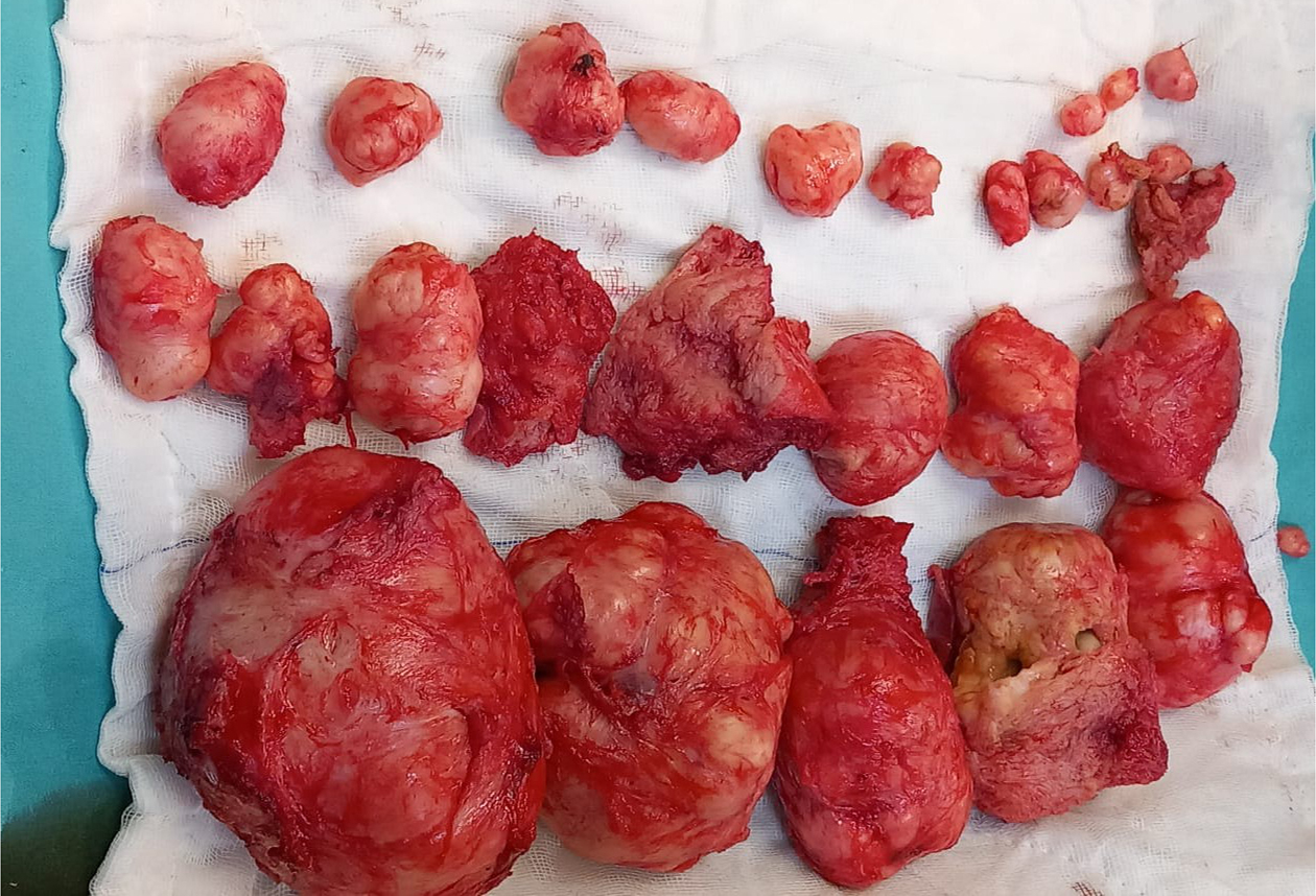
Myomas removed from our patient during the operation performed while preserving the uterus
Myoma Treatment
Myomas are treated or only monitored by considering their size, number, problems they cause, and imaging features. For myomas that are small in diameter, few in number, embedded in the uterine wall or growing outward, not causing problems such as bleeding, pain, infertility, miscarriage, etc., and not growing excessively during follow-up, monitoring alone without any treatment may be sufficient. For larger myomas that cause problems such as bleeding, pain, infertility, miscarriage, or premature birth, planning a treatment would be the right approach.
There are multiple treatment options for myomas. There are many treatment options available to reduce bleeding in myomas that are thought to only cause irregular bleeding and in which no pathology is detected in the pathological examination of samples taken from the uterus (endometrium) due to this irregular bleeding, from non-steroidal anti-inflammatory drugs to intrauterine hormonal IUDs and oral hormones. The important point here is that it is absolutely certain that there are no abnormal cells in the biopsies taken from the uterus. Because, in addition to the myoma, the patient may also have thickening (endometrial hyperplasia), polyps or cancer in the uterus at the same time. If bleeding is attributed to the myoma, and no samples are taken from the endometrium, it may cause a delay in the treatment.
Surgical treatment for myomas is applied to large-scale myomas, those suspected of cancer, those causing reproductive problems, or in cases where the desired response cannot be obtained with medical treatment. In this case, options such as myoma removal or complete removal of the uterus will be considered, considering the patient's age, whether there is a reproductive expectation, etc. Both myoma surgeries (myomectomy, fibroidectomy: removal of the myomas)) and hysterectomy (removal of the uterus) can be performed openly (laparotomy) or closed (laparoscopy or hysteroscopy). When choosing the surgical method, it will be necessary to consider which method will be most possible, easiest, and least traumatic to remove the myoma. For example, while a small myoma that has grown into the inner part of the uterus (endometrium) can be removed with hysteroscopy (hysteroscopy is explained under a separate heading and is the procedure of imaging the inside of the uterus by passing through the vagina and cervix with the help of a camera without an incision), hysteroscopy will not be appropriate for surgery for a myoma that has not been connected to the inner part of the uterus or has grown outward.
Abdominal myomectomy, that is, laparoscopy or laparotomy (entering the abdomen with an incision similar to a cesarean section), are also methods used to remove myomas.
Laparoscopic Myoma Surgery
Laparoscopy is a procedure performed by entering the abdomen through a small incision with a camera. It is performed under anesthesia in operating room conditions. In order to remove the myomas, in addition to a camera incision, several incisions are made for the entry of instruments. The abdomen is inflated with carbon dioxide and the myomas are removed from the uterus. Since it is not possible to remove the myomas through small incisions in laparoscopy myoma surgery, they will need to be fragmented (morcellation). As literature information accumulates that if there are cancer cells in the myoma during the fragmentation procedure, these cells spread into the abdomen, laparoscopic myoma surgeries have begun to be viewed with more caution.
Myoma Surgery with Laparotomy (Entering the Abdomen with an Incision)
In laparotomy myoma surgery, the abdomen is usually entered through an incision similar to a cesarean section, the myomas are seen and removed without being broken down. In this procedure, the possible spread of cancer cells is not expected like in laparoscopy and morcellation, but the patient's return to daily life after the surgery will be a few days longer than with laparoscopy.
Some procedures such as blocking the myoma vessels (embolization) or reducing the volume of myomas with improved ultrasonographic modalities have also been described today for the treatment of myomas. However, gynecologists generally stay away from such myoma reduction methods because their aim is to completely remove the myomas and send them to pathology (to rule out cancer).