






Posterior Urethral Valve (PUV)
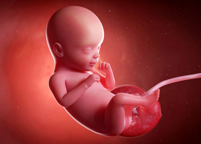
Posterior Urethral Valve (PUV) is a developmental disorder affecting approximately 2 out of every 10,000 fetuses diagnosed in the womb. In fetuses, urine cannot exit the bladder due to the formation of valves (or small valves) in the posterior urethra, the first exit point of the urethra. Urine accumulates there, and as this accumulation progresses, the ureters (the channels that carry urine from the kidneys) and the renal pelvis (the area where urine first collects in the kidneys) enlarge. As the accumulation continues, it begins to damage the kidneys, leading to increased echogenicity, kidney cysts, and ultimately, kidney dysfunction. Urine accumulating in the bladder can cause an increase in pressure, leading to bladder rupture and leakage of urine into the baby's abdomen (fetal urinary ascites).
Due to the inability of urine to exit the bladder, the baby's amniotic fluid gradually decreases. A decrease in amniotic fluid is a problem that reduces the baby's chances of survival both in the womb and after birth, and carries a risk of death. Amniotic fluid is vital for the development of the baby's lungs. In cases where there is little or no amniotic fluid, the baby's lung development does not occur as desired, and babies face the risk of dying from lung failure after birth. In cases of decreased amniotic fluid, the umbilical cord may also be compressed, and the baby may lose its life in the womb.
As can be easily understood from the above explanation, most PUV cases (approximately three-quarters in the literature) are lost if not treated in utero. However, in babies who undergo surgery in utero, the chance of survival can reach 75 percent.
How is Posterior Urethral Valve (PUV) Diagnosed?
Posterior Urethral Valve (PUV) can be easily diagnosed during fetal examination using ultrasonography after the first trimester of pregnancy. Since the kidneys play a limited role in amniotic fluid formation during the first trimester, diagnosis is usually made after the 12th week. During ultrasonography, the baby's bladder appears larger than normal, the urethra (the exit point of the bladder) is widened (keyhole sign), the bladder wall is thickened, the area where urine first collects in the kidneys and the ducts carrying urine from the kidneys to the bladder are widened, and in more advanced cases, the kidney tissue appears brighter (hyperechoic kidney) and renal cysts are present in the kidneys. Therefore, Posterior Urethral Valve (PUV) in these cases can be diagnosed with a careful ultrasound examination in the womb. We also use the criteria mentioned above for evaluation in our clinic.
How is the Decision Made for Intrauterine Surgery in Cases of Posterior Urethral Valve (PUV)?
In cases of Posterior Urethral Valve (PUV), intrauterine surgery is now primarily performed not to protect the baby's kidneys, but to help them survive. Since infants with low amniotic fluid levels experience negative lung development and are often lost due to pulmonary hypoplasia, the primary goal of intrauterine surgery is to prevent further amniotic fluid deficiency, thus aiding lung development and ultimately survival of the newborn. As these statements indicate, infants with enlarged bladders and reduced amniotic fluid are expected to benefit from intrauterine surgery and are candidates for the procedure. Due to the associated risks, surgery is approached with more caution in cases where amniotic fluid is not reduced. Before performing intrauterine surgery, a urine sample is taken from the fetus's bladder (vesicocentesis) to analyze the fetus's urine and obtain information about kidney function. However, since a robust test to accurately assess kidney function has not yet been found, this practice is now questioned. At the Delhi roundtable meeting, routine urine analysis from infants using urine samples was not recommended. The suggestion to evaluate whether the bladder is full after emptying it via vesicocentesis was put forward. Since intrauterine surgery is primarily performed to contribute to the infant's survival and not to prevent long-term kidney damage, dialysis, or the need for kidney transplantation, this recommendation seems reasonable and consistent with current scientific data.
How is Intrauterine Surgery Performed in Cases of Posterior Urethral Valve (PUV)?
There are essentially two different intrauterine surgical methods for Posterior Urethral Valve (PUV) cases:
- Vesicoamniotic Shunt (VAS)
- Fetal Cystoscopy and Laser
Vesicoamniotic Shunt (VAS)
A vesicoamniotic shunt (VAS) is a flexible catheter designed to drain urine from the baby's bladder into the amniotic cavity. One end of the catheter is placed in the baby's bladder, and the other end remains outside the baby's skin. By draining the urine accumulated in the bladder into the amniotic sac, it both empties the baby's bladder and prevents a decrease in amniotic fluid, contributing to the baby's lung development. The procedure is usually performed under ultrasound guidance after the mother is given local, regional, or general anesthesia, and the baby is given anesthesia and muscle relaxants (via the umbilical cord or extremity). It is a short procedure with minimal risks to the mother. However, it carries the potential for complications such as procedural failure, rupture of membranes after the procedure, premature birth, shunt blockage, and shunt displacement, thus requiring the insertion of multiple shunts. Current literature shows that VAS procedures increase the survival chances of babies. However, data on preventing long-term kidney damage is limited.
Fetal Cystoscopy and Laser
Fetal cystoscopy and laser surgery are among the areas where current advanced technology allows for use in medicine. During the procedure, after the mother is given anesthesia (local or general), the fetus in the womb is also given anesthesia and muscle relaxants. This ensures anesthesia for the baby and prevents movement during the procedure. The system, called fetoscopy, is guided into the fetus's bladder under ultrasound guidance. The inside of the bladder is evaluated with a camera, and it is determined whether the problem at the bladder outlet is PUV or urethral stenosis or atresia. After the diagnosis of posterior urethral valve is confirmed, the valve area is opened with a laser, providing a passage for urine flow. Fetoscopic laser valve ablation can provide definitive treatment for posterior urethral valve in the womb. However, complications such as procedural failure or damage to surrounding tissues have also been reported in the literature. In our cases, both the success of the procedure and the complications related to the procedure are similar to those in the literature. There is promising data supporting the idea that fetoscopic laser surgery offers a greater chance of kidney preservation after birth compared to vesicoamniotic shunting. However, the relative novelty of the procedure and the limited data on long-term outcomes are factors that need to be considered in patient selection and education.
In conclusion, in carefully selected cases of posterior urethral valve patients, intrauterine surgery increases the survival rate of the infant. More data is needed regarding its positive long-term effects on kidney function.